Dermatological Infections: Common Pitfalls to Dodge

By: Dr Ian H Coulson,
DermNet Editor-in-Chief and Consultant Dermatologist
Lancashire, United Kingdom.
Whenever my defence organisation periodical drops into my inbox, it is a priority to open it. It is always instructive to read of cases where things have gone wrong. So frequently, there is a ”there but for the grace of God” moment.
On this page, I hope to cover some of the more common mistakes that are made (and indeed I have made) within the practice of dermatology, in the hope that others may benefit from our discomfort!
Maxims will be interspersed within the text that are always worth reflecting on.
“More mistakes are made in dermatology by not looking, rather than not knowing”
Blunders with bacteria!
With so much of dermatology concentrating on control rather than cure, curable bacterial infections need our utmost concentration.
Case 1:
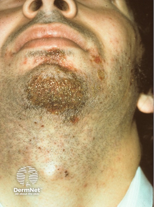
This man has a weepy rash on his chin which started four days ago. A putative diagnosis of impetigo was made on the basis of its short duration and the typical honey-coloured crust. As it is extending, despite the use of topical fusidic acid, the diagnosis is now questioned.
If there is ever a question of the diagnosis of impetigo, take a skin swab. In this case, it showed a fusidic acid-resistant strain of Staphylococcus aureus. In some regions where topical fusidic acid use is high, then so are resistant strains. Erythromycin resistance is more common than mupirocin resistance, and more common than MRSA. If in doubt, treatment should be guided by culture sensitivities.
Case 2:
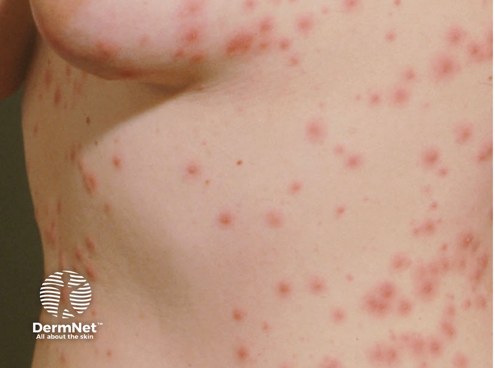
This young woman has developed follicular pustules over her trunk. It is not responding to flucloxacillin, so correctly, a skin swab was taken.
It has grown Pseudomonas, and she was embarrassed to disclose a “skinny dip session” in a hot tub with a few others, now similarly affected. Remember that folliculitis can be sterile, as well as being caused by gram-positive and negative bacteria, as well as dermatophytes and yeasts.
Not all pustules are pyococcal!
Case 3:
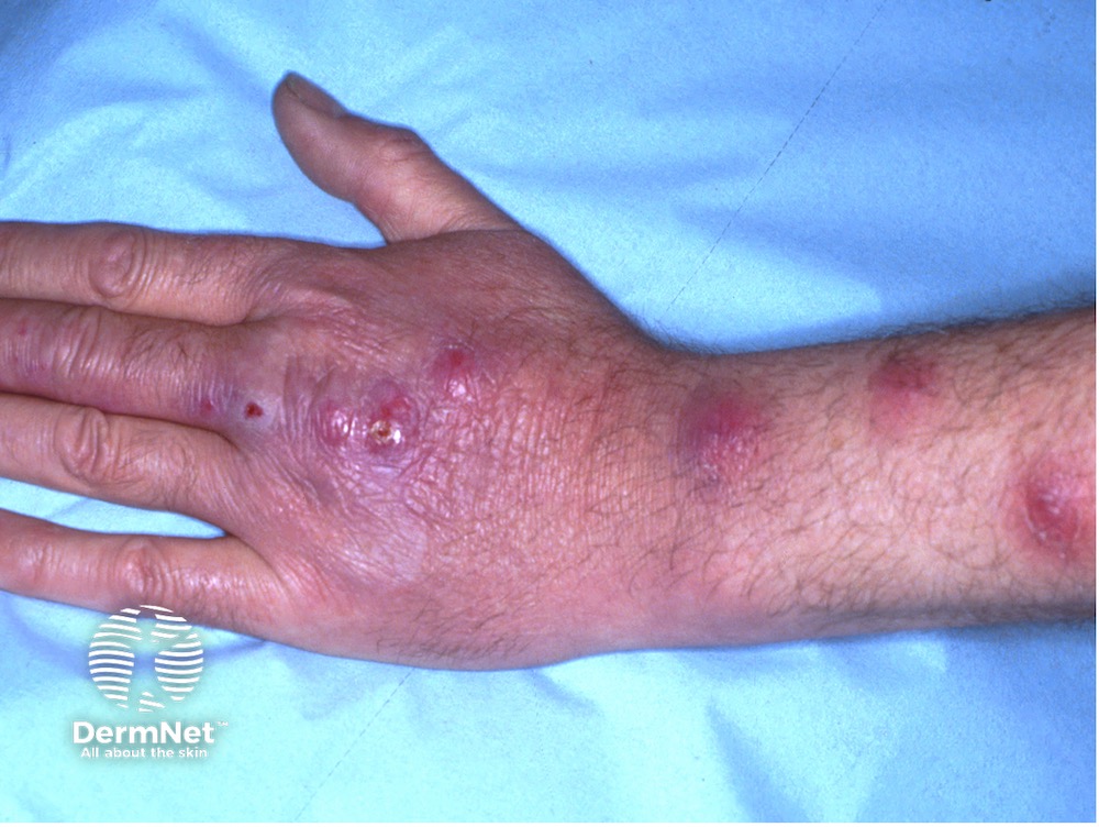
This man developed a tender papule on his ring finger, and similar lesions have developed more proximally over the last 6 weeks. They are now beginning to suppurate; a skin swab grew only normal commensals. It has not responded to oral augmentin. He denies keeping fish.
On culture of biopsy specimens, and specifically asking for a low temperature culture, Mycobacterium marinum was grown, and an IRGA TB test was positive. Mycobacteria will not grow on conventional culture material, and the lab needs to be primed so that material, ideally tissue, is cultured on the correct medium. A forensic history revealed that he was a skilled odd job man, and he’d fixed a neighbour’s tropical fish tank aerator pump. So he was right to deny keeping fish, but his neighbours’ fish were the source of his infection!
Case 4:
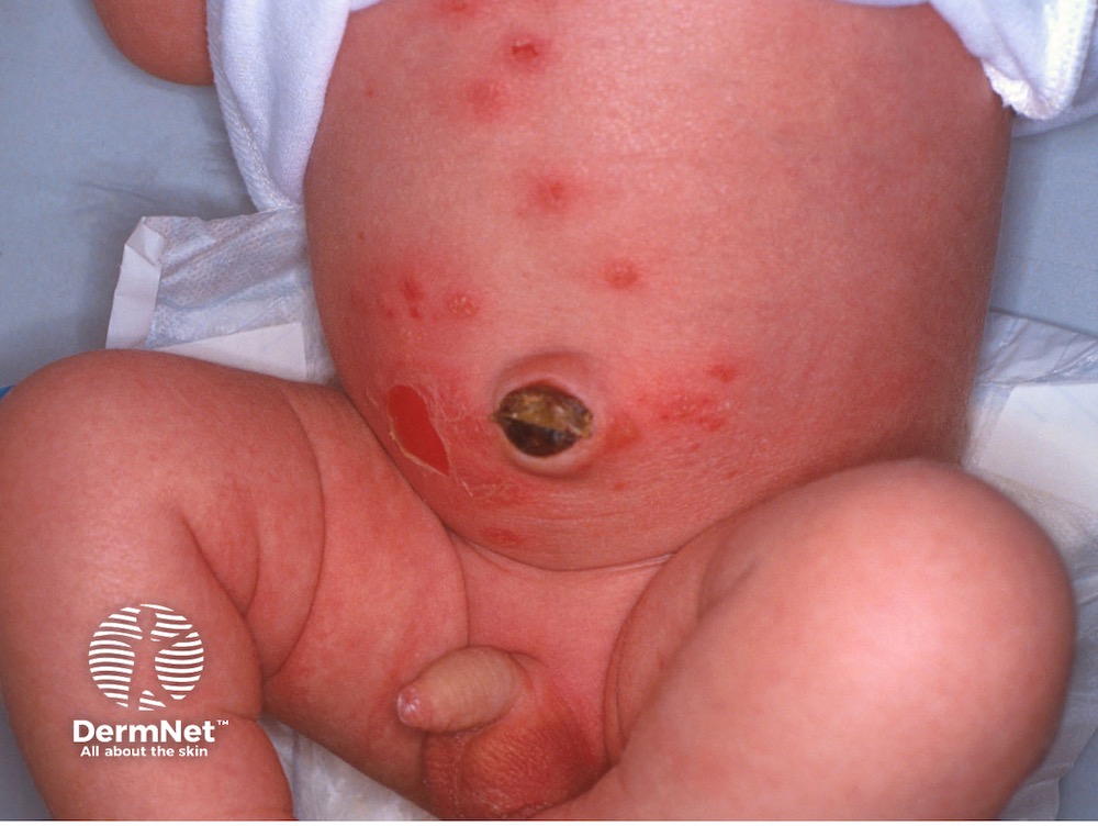
This boy is five days old, and he has developed blisters over his lower abdomen. His parents and his neonatologist have already consulted Dr Google! Everyone is fearing that this is epidermolysis bullosa.
Skin swabs from the blisters and the umbilicus have grown Staphylococcus aureus – and the blistering resolved rapidly with the introduction of anti-staphylococcal antibiotics. A great relief.
Remember that impetigo, caused by the right phage type of staphylococcus, can blister.
Case 5:
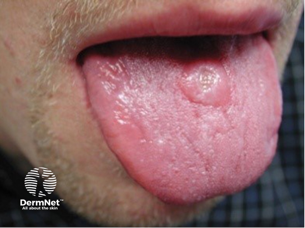
This man in his 20s has developed an ulcer on his tongue. He is otherwise well. Swabs for bacteria and herpes simplex have proved negative.
The diagnostic test was a PCR for Treponema pallidum – it was positive. He has primary syphilis, and over 30% of primary syphilitic chancres are now either intraoral or on the lips, as unprotected oral sex in both the heterosexual and MSM populations is widespread.
Faults with fungi
“A unilateral or asymmetric scaly rash is tinea ‘till proven otherwise!”
Case 6:
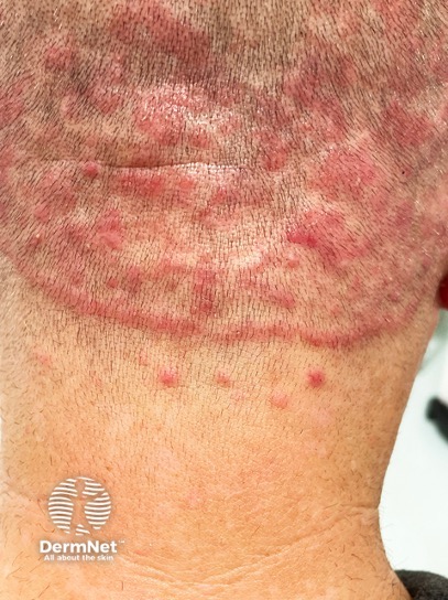
This man has a spreading rash over his scalp. He has a past history of hand eczema, and patch testing showed he was allergic to methylisothiazolinone and perfumes. He has been applying some of his topical steroid to the scalp – it has helped the itch but made the rash spread. A course of flucloxacillin has not helped.
This is tinea incognito in an unusual site – a fungal infection modified by the use of topical steroids. The modification is the extension and also the development of follicular pustules that have mimicked a bacterial folliculitis. Note also the obvious “edge” around his neck. The steroid allows the fungus to invade the follicles, inducing a fungal folliculitis.
Look elsewhere; take his socks and shoes off – he has interdigital and moccasin type tinea pedis and toenail tinea unguium. Scrapings from the sole and scalp grew Trichophyton rubrum. Oral terbinafine came to the rescue!
Case 7:
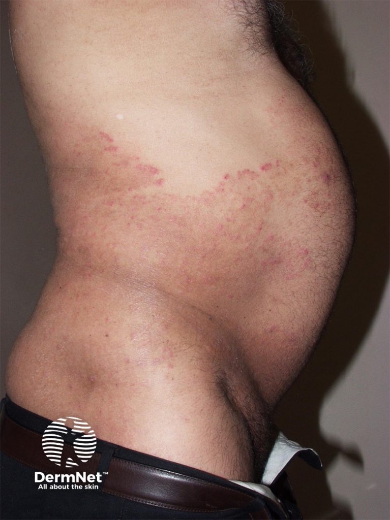
This man has had a spreading eruption over his groin and abdomen. Mycology has demonstrated fungal hyphae; the culture grew Trichophyton mentagrophytes. Oral terbinafine has been exhibited for 2 months and the eruption has spread despite this. What is going on?
The patient’s skin type should be a clue – he is a UK resident but visits India and Pakistan regularly on business. Therapy-resistant dermatophyte infection is now commonplace in the Indian subcontinent and spreading globally. Strains may be resistant to allylamines, azoles or both. The causative strains are often reported as T mentagrophytes, but this may be confused with the newly identified therapy-resistant T. indotineae. Treatment is difficult and itraconazole is more likely to produce a therapeutic response.
Combination of oral antifungal agents plus topical antifungal agents such as azoles, ciclopirox or amorolfine may improve responses.
Case 8:
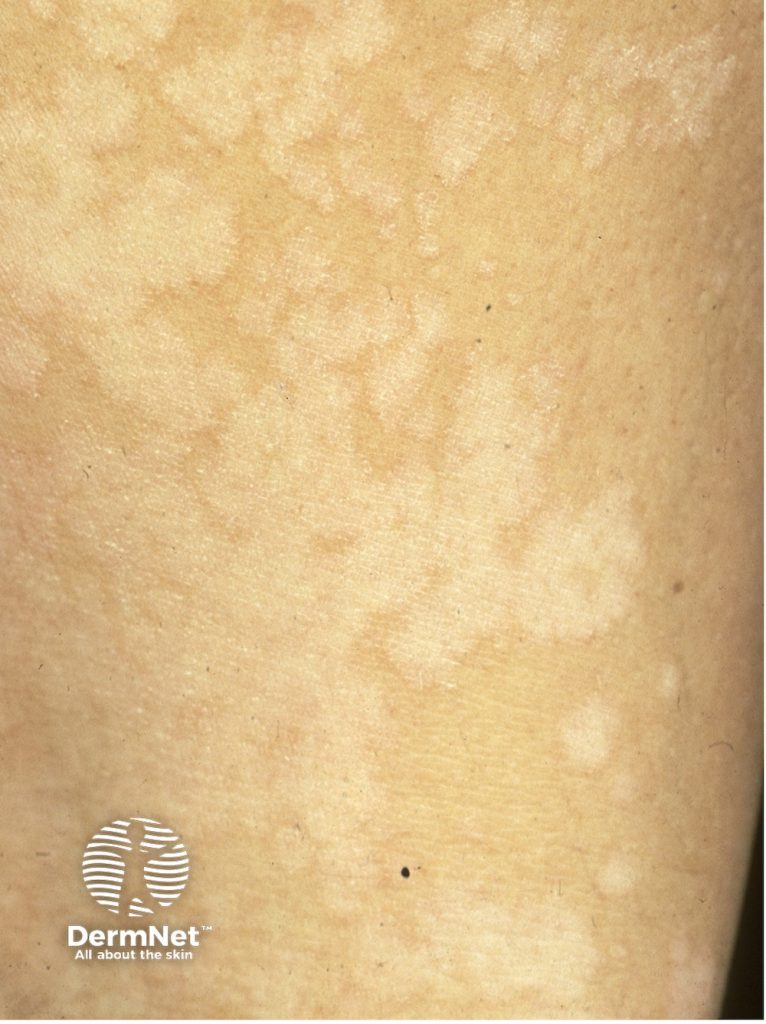
This 18-year-old has this scaly rash over his chest. It has been diagnosed as a “fungal infection” but has not responded to oral terbinafine. The diagnosis is now being questioned!
This is typical Pityriasis versicolor, it is easy to confirm on direct mycology by the recognition of the pseudohyphae and spores (meatballs and spaghetti), although the causative pityrosporum yeast requires special media to culture it.
Although it may be sensitive to topical terbinafine, this is an inefficient and expensive topical option, and oral terbinafine is not effective. Selenium sulphide or ketoconazole shampoos are more effective and economical. Short courses of oral itraconazole are often patient preferred.
Remember to let the patients know that the depigmentation that will develop due to the causative yeast releasing azelaic acid will prevent melanogenesis for up to 6 months and this is not the result of persisting infection. The presence of bran-like light brown scale however does suggest either recurrence or reinfection.
Case 9:
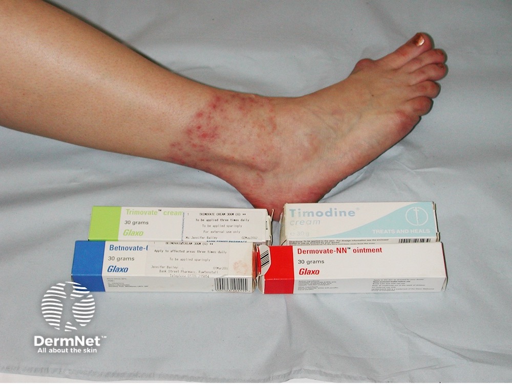
This lady has a continuing problem on her ankle that has persisted despite antifungal-containing creams. What is the mistake?
Again this is tinea incognito; all of the agents contain steroids of differing potency. Nystatin is present in Dermovate NN, Timodine and Trimovate, and this is active only against yeasts rather than the causative dermatophyte fungi. Look between the toe webs and take her nail varnish off – she has tinea pedis and unguium and will require a systemic antifungal.
“Experience – making the mistakes with greater confidence!” So keep on your toes!
For more dermatology cases that can be expected in the real world, see DermNet Case Library.